
Nearby in the archive
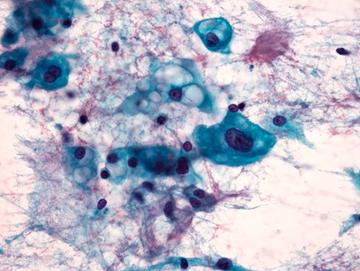
Papanicolaou stained smear of a C2 vertebral chordomal mass, microscopy. Chordomas are cancers formed of cells which resemble those of the notochord (spine) of a developing foetus. Although they can present anywhere within the spine and skull, the majority grow in the sacral region of the spine, corresponding to the lower back. This image shows a Papanicolaou (pap) stained smear obtained from a needle biopsy of a chordoma of the C2 vertebrae, located at the top of the neck just underneath the base of the skull.
William R. Geddie
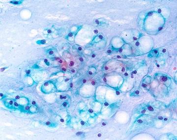
Papanicolaou stained smear of a clival chordoma, microscopy. Chordomas are cancers formed of cells which resemble those of the notochord (spine) of a developing foetus. Although they can present anywhere within the spine and skull, the majority grow in the sacral region of the spine, corresponding to the lower back. This image shows a Papanicolaou (Pap) stained smear obtained from a needle biopsy of a chordoma in the clivus, a part of the cranium at the base of the skull.
William R. Geddie

Parotid Cyst amylase crystals 1
William R. Geddie
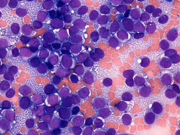
PNET MGG 63x 2
William R. Geddie
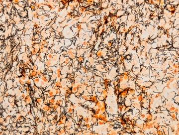
Spirochete presentation in inguinal lymph node section
William R. Geddie
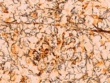
Spirochete presentation in inguinal lymph node section
William R. Geddie
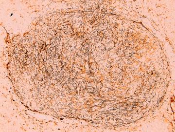
Spirochete presentation in inguinal lymph node section
William R. Geddie
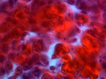
Squamous carcinoma of lacrimal gland, microscopy
William R. Geddie